|
|
|
|
|
|
|
|
Iuliana Morariu,andrew dias,Aongus Curran
|
|
|
Ir Med J. 2012 Jun;105(6):184-5
I Morariu, A Dias, A Curran
Royal Victoria Eye & Ear Hospital, Adelaide Road, Dublin 2
Abstract
We report a case of a large pleomorphic adenoma arising from the deep lobe of the parotid gland in the parapharyngeal space and causing obstructive sleep apnoea. Review of literature, clinical features, pathology, radiological findings and treatment of these tumours are discussed.
|
|
Introduction
Tumors in the parapharyngeal space are relatively rare and constitute less than 0,5% of head and neck neoplasm1-4. Of these, pleomorphic adenoma is the commonest benign tumour (40%)1. Most tumours originate in the superficial lobe of parotid gland but, more rarely, these tumours may involve the deep lobe of the parotid gland.3,5 The main characteristics are a high recurrence rate and infrequent malignant conversion.6
Case Report
A 42-year-old man of Asian origin, with no previous clinical history of note, was referred to our Emergency Department with pharyngodynia, change in quality of speech, heavy snoring and sleep apnoea of one-year duration. Physical examination showed a huge, smooth, firm swelling on the right side of the soft palate and right lateral pharyngeal wall, with a normal overlying mucosa, with medial displacement of the soft palate and uvula to the left, causing obstruction to the airway. There was no significant lymph node enlargement in the neck. Bimanual palpation of the right parotid gland revealed no abnormalities. Flexible nasofibroscopy showed extension of swelling into the nasopharynx with very narrow airway on this level. The lower limit of swelling was at the level of vallecula. At admission, the patient demonstrated symptoms and signs of obstructive sleep apnoea, namely unexplained daytime sleepiness, loud snoring with periods of silence followed by gasps and reduction in blood oxygen saturation up to 70% in supine position on pulse oximetry.
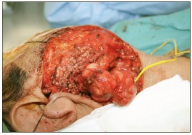
Magnetic resonance imaging demonstrated a 7×6×4 cm mass arising in the right parapharyngeal space. The lesion appeared to be in continuity with the deep lobe of the parotid gland. It contained internal septation with fluid components and was circumscribed. The lesion demonstrated a considerable mass effect, with significant narrowing of the oropharynx. A CT Angiogram of the carotid arteries showed a large right oropharyngeal mass which splayed the right internal and external carotid arteries, without any abnormal vascularity. Fine Needle Aspiration Cytology, performed transorally, revealed cytological caracteristics of a pleomorphic adenoma. Complete excision of the lesion was performed via a cervical transparotid approach with preservation of the facial nerve, after informed consent. At surgery, the tumour was found to be arising from the deep lobe of the parotid gland. Histopathology confirmed the mass to be a pleomorphic adenoma with complete excision. The post-operative period passed without incident and marked improvements in nocturnal hypoxic episodes and the symptoms of obstructive sleep apnoea were seen, with no recurrence at six month.
Figure 1: Bulging of the tumour in the right soft palate, with medial displacement of the
uvula to the left with significant narrowing of the oropharynx
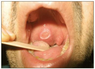
Figure 2: Intra-operative clinical photograph of surgical excision of the tumour
Discussion
Parapharyngeal space tumours are infrequent and usually benign. They present with minimal symptoms, hence early diagnosis is difficult. Few reports describe obstructive sleep apnoea symptoms secondary to parapharyngeal space lesion8. Pleomorphic adenomas frequently present as a painless firm mass. In most cases they do not cause ulceration of the overlying mucosa7,9. Generally they are mobile, except in the hard palate. As tumours enlarge, they may compress the lower cranial nerves, causing hoarseness, dysphagia, dysarthria or Horner’s syndrome3,5. Diagnostic imaging, namely computed tomography or magnetic resonance imaging are mandatory. Magnetic Resonance Imaging is preferred, because of better definition of soft tissue. It demonstrates precise information regarding tumour margins and tumour relationship with the surrounding structures.3 Angiography is indicated when carotid artery involvement is suspected or in paragangliomas and highly vascular tumours10. Fine Needle Aspiration Cytology is indicated following diagnostic imaging in order to exclude a vascular lesion. Open neck or trans-oral biopsies are best avoided, as incising the tumour capsule increases the risk of recurrence.3 These tumours should be considered in the differential diagnosis of common infectious or traumatic conditions9.
The treatment of pleomorphic adenoma is essentially surgical. Various approaches have been described in the literature2-9. The cervical-trans-parotid approach is the most frequently used approach as it allows excision of benign lesions with good control of vascular and nervous structures following dissection of the superficial parotid lobe. Postoperative radiotherapy to the parapharyngeal space could possibly reduce the recurrence rate in such tumors2. After excision of the tumour, clinical evaluation and imaging studies should be performed yearly or biannually to out-rule recurrences, which can occur up to 20 years5.
Correspondence: I Morariu
16 Cloghanboy Ave, Ballymahon Road, Athlone, Co Westmeath
Email: [email protected]
References
1. Ladeinde AL, Adeyemo WL, Concurrent pleomorphic adenoma in parapharyngeal space and submandibular gland. World Journal of Surgical Oncology 2004, 2:6
2. Varghese BT, Sebastian P, Pleomorphic adenona of minor salivary gland in the parapharyngeal space. World Journal of Surgical Oncology 2003, 1:2
3. Sergi B, Limongelli A, Giant deep lobe parotid gland pleomorphic adenoma involving the parapharyngeal space. Report of three cases and review of the diagnostic and therapeutic approaches. Acta Otolaryngologica Italica 2008, 28:261-265
4. Spiro RH. Salivary Neoplasms: overview of a 35 year experience with 2807 patients. Head Neck Surgery. 1986;8:177–184
5. Waldron CA, El-Mofty SK, Gnepp DR. Tumours of the intra oral salivary glands: A demographic and histologic study of 426 cases. Oral Surg Oral Med Oral Pathol. 1988;66:323–333
6. Ferro MF, Sanroman JF, Surgical treatment of benign parapharyngeal space tumours. \presentation of two clinical cases and revision of the literature. Med Oral Patol Oral Cir Bucal. 2008 1:13:E61-4
7. Clauser L, Mandrioli S, Pleomorphic adenoma of the palate. J Craniofac Surg 2004;15:1026-9
8. Eveson JW, Cawson RA. Tumour of the minor (oropharyngeal) salivary glands: demographic study of 336 cases. J Oral Pathol. 1985;14:500–509
9. Moraitis D, Papakostas K, Pleomorphic adenoma causing acute airway obstruction. Journal of Laryngology & Otology 2000, 114:634-636
10. Giddings CE, Bray D, Pleomorphic adenoma and severe obstructive sleep apnoea. Journal of Laryngology &Otology 2005, 119:226-229
|
|
|
|
Author's Correspondence
|
|
No Author Comments
|
|
|
Acknowledgement
|
|
No Acknowledgement
|
|
|
Other References
|
|
No Other References
|
|
|
|
|